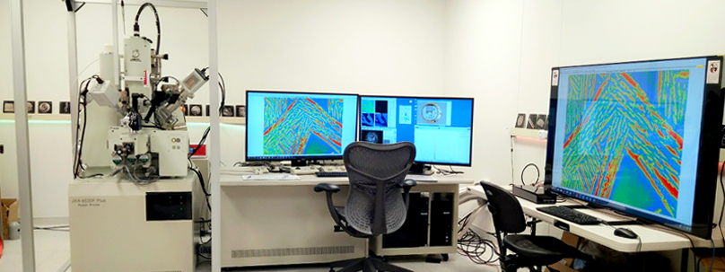
I am managing the electron microprobe lab of the University of Minnesota that offers non-destructive chemical analyses of solids. The electron microprobe is capable of quantitatively measuring the abundance of all elements from B to U and combines micron-scale chemical analyses with scanning electron microscopy, capable of large- and small-scale element mapping of specimens.
The new lab in Tate Hall houses a new state-of-the-art JEOL JXA-8530FPlus Electron Probe Microanalyzer (EPMA) with four wavelength-dispersive spectrometers (WDS), SDD-EDS and SEI, BSE and CL imaging capabilities. A unique feature of the system is a novel Soft X-Ray Emission Spectrometer "SXES" as the fifth spectrometer, the first in an academic institution in North America.
The SXES spectrometer uses X-ray focusing optics, variable line-spacing gratings and a solid-state X-ray CCD detector to collect a low energy X-ray spectrum. It provides higher spectral resolution and higher sensitivity for soft X-rays (<1 keV) relative to traditional WDS, yielding information on the electronic structure and bonding [1-2]. The grating and CCD detector are rigidly fixed, with no moving parts, resulting in spectra that are extremely reproducible.
A characteristic feature of the SXES is its parallel detection of the X-ray signal. This allows it to be used like a conventional energy dispersive spectrometer (EDS) and generate a spectrum without the need to move the grating or the detector (like a traditional WDS scan). Hyper-spectral data is collected simultaneously, like an SDD EDS, with orders of magnitude better energy resolution. This parallel collection of the spectra data allows for the acquisition of spectra maps, from which element maps and chemical state maps can be extracted.
The SXES spectrometer at the University of Minnesota is configured with one high-resolution (JS300N) and one broad energy range (JS2000) grating with a combined spectral range covering nominally the X-ray energy range of 85 eV through >2.5keV. This allows for the analysis of ultra-light elements with very high peak to background ratios (the spectrometer is capable of trace analysis of light elements such as C, N and B in steel at the 10-100ppm range), but also a wider element list using both first order and high order reflections including the first order L-lines of the transition elements. For example, it is possible to study iron oxidation states from the peak shape of Fe-L spectra.
[1] H. Takahashi et al. 2014 JEOL News 49 73-80.
[2] M. Terauchi 2014 In: Transmission Electron Microscopy Characterization of Nanomaterials, 287-331. (Springer: Berlin, Heidelberg).
More information can be found here.
